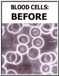
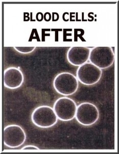
 Darkfield microscopy is a special type of photography that makes the blood look like the images you see here. As seen in the picture on the left, darkfield microscopy reveals the toxins in the blood and blood cells as cloudiness. These toxins can consist of anything that a phagocytic cell would ingest. The after picture on the right was taken after the dermal chelation therapy process was completed. It shows that the cloudiness is gone from both the cells and the blood. This indicates that the toxins, THMs if present, are no longer in the cells or blood.
Dermal Chelation Therapy and Chronic Auto-Immune Disorders
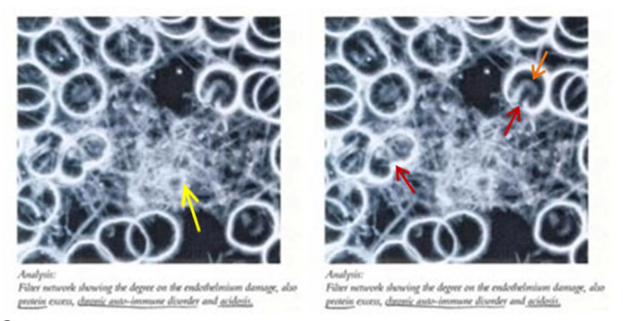
Below are some pictures taken from the same research. The same darkfield microscopy image of an individual’s blood with a chronic autoimmune disorder is shown twice. The left picture below highlights the excessive amount of protein (yellow arrow) in the blood due to a chronic autoimmune disorder. The picture is repeated with orange and red arrows pointing to the misshapen cells. Notice how dim the white of the outer membrane becomes at the areas of severe damage.
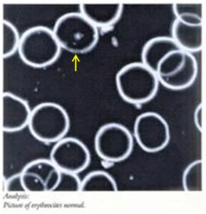
The picture on the left ([2]) shows the blood cells of the same person with the chronic autoimmune disorder after the dermal chelation process. The excessive amount of protein is no longer present. The blood cells appear nearly normal except for where there is still some occasional dimming of the outer membrane.
Dermal Chelation Therapy and Chronic Auto-Immune Disorders Lacking Oxygen
According to the same research ([3]), the picture below on the right side shows blood lacking in oxygen, in an acid state and developing antibodies to its own tissue, as is often seen with chronic autoimmune disorders. Note the resulting stacking of the blood cells.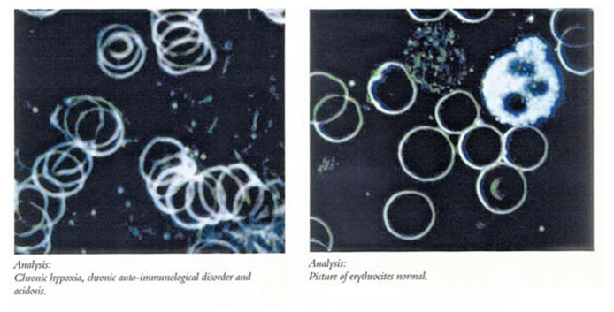
Since the dermal chelation therapy process allows oxygen to enter through the pores of the feet and eventually reach the blood, the blood cells become oxygenated. Note that after the dermal chelation process is performed, there is no stacking of the blood cells as seen in the picture to the right above. The blood cells appear to be almost normal.
As we have shown in the figures of resulting dermal chelation therapy water above and in agreement with our preferred equipment manufacturer’s research results presented above, the resulting water looks very different from one person to the next. The dark microscopy pictures of the blood make it apparent that our preferred equipment is interacting with the body. We hope it is obvious at this point that, with our preferred equipment, there does appear to be specific induction of toxic elements released through the feet.
Next:
Resulting Dermal Chelation Therapy Water Containing Ammonia, Nitrates, and Phosphates
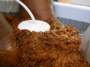
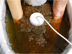
Understanding Colors and Textures of the Resulting Dermal Chelation Therapy Water that Indicates:
Understanding Colors and Textures of the Resulting Dermal Chelation Water that Indicates:
| Bubbly Foam | Dark Green | Blood | Nicotine | Yellow-green |
|---|---|---|---|---|
 |  |  |  | 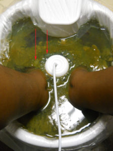 |

Popular Topics
Optimum Health’s Concept of Healing
Primary Wellness Consultations
Email Us: How Can We Assist You?
 Start your path to optimum health by scheduling your appointment today. Member: Certified Natural Health Professionals


References:
[1] Aqua Detox. Scientific Research, The Evidence. 2008;8
[2] Aqua Detox. Scientific Research, The Evidence. 2008;11
[3] Aqua Detox. Scientific Research, The Evidence. 2008;12