FIGURE 1
A concentration of 2.5 µg/ml of silver nanoparticles (Ag-NPs) stimulates the body to increase stem cell production (shown in units of IL-8) by over 200%. [1] A concentration of 1 µg/ml of silver nanoparticles (Ag-NPs) and silver ions (AG-ions) stimulate the body to increase stem cell production by a little over 100%. Therefore, AG-NPs are best for stimulating stem cell production.
[1] Greulich, C., Kittler, S., Epple, M. et al. Studies on the biocompatibility and the interaction of silver nanoparticles with human mesenchymal stem cells (hMSCs), Langenbecks Arch Surg. 2009 Natl 394(3): 495-502. Doi:10.1007/s00423-009-0472-1. Epub 2009 Mar 12.

FIGURE 2
This is the proposed mechanism for Silver nanoparticles (AgNPs) inhibiting the cycling, growth and survival of cancerous epithelial cells. This shows that silver can have an affect on cells. The schematic diagram represents the possible cellular uptake of silver nanoparticles by active and passive processes in eukaryotic cells. AP-2, Adaptor complex; PH domain, pleckstrin homology domain; PKB/Akt, Protein kinase B or Akt; PI3K, phosphatidylinositide 3-kinases; Rab5, Ras related protein; APPL1, a4 precursor protein like 1; EEA1, early endosome-associated protein.[2] We, at Optimum Health, have found that silver has the opposite affect on stem cells at all three levels.
[2] https://www.mdpi.com/1422-0067/17/10/1603/htm
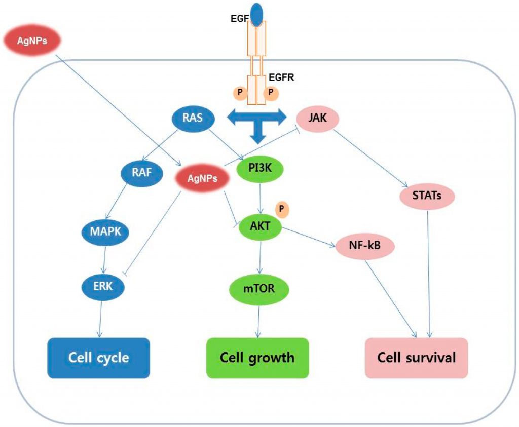
FIGURE 3
Ample supplies of vitamin C are required to make and maintain the ground substance, an amorphous gelatinous matrix that acts as the glue that holds cells together in bodily structures like arteries, ligaments, tendons, muscles and others.[3] Without vitamin C, the ground substance weakens and the cells are not able to maintain proper structure allowing micarovascular leakage.[4]
[3] https://slideplayer.com/slide/13206852/
[4] Barbara, get primal panacea book from my purse or my office and enter it here.
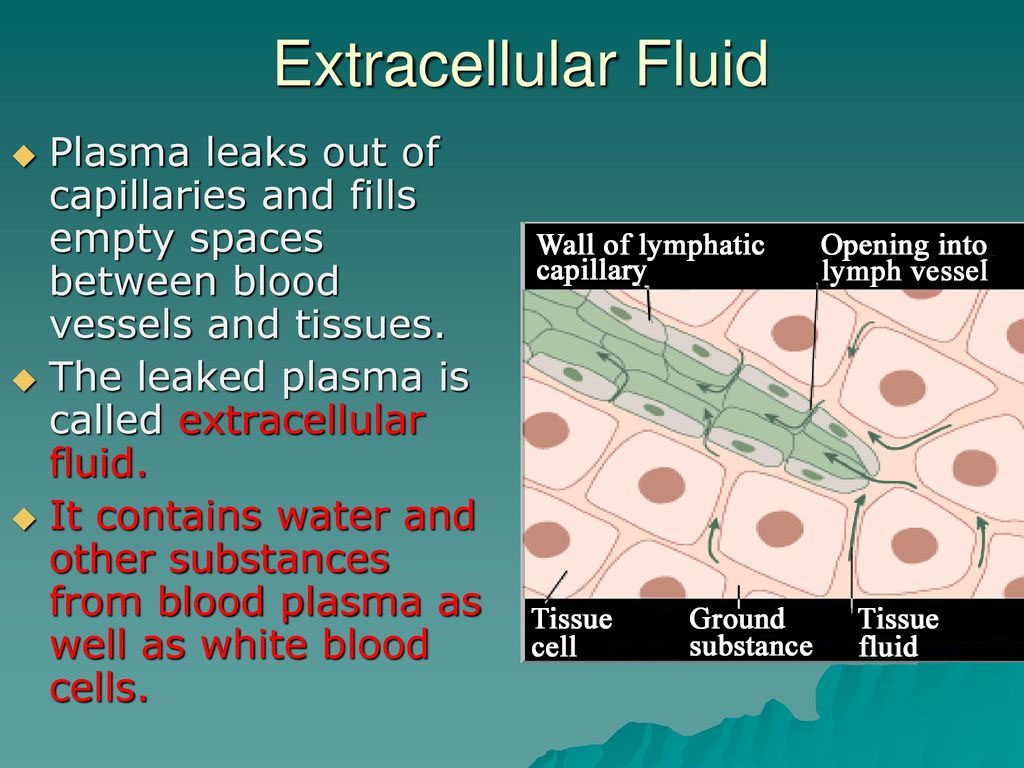
FIGURE 4
Hand caught on fire when he was trying to put out a grease fire. Note that the increase in microvascular leakage has caused the burn site to become inflamed and weepy.
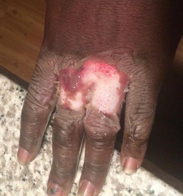
FIGURE 5
Microvascular leakage continues. Lack of professional attention has allowed the infection to go unchecked.

FIGURE 6
Vitamin C has been initiated. Microvascular leakage is no longer an issue, therefore, the burn site has dried out. Notice how the skin has begun to peel off on the distal end of the middle digit. This is what we see when the colloidal silver stimulating the regeneration of the tissue.
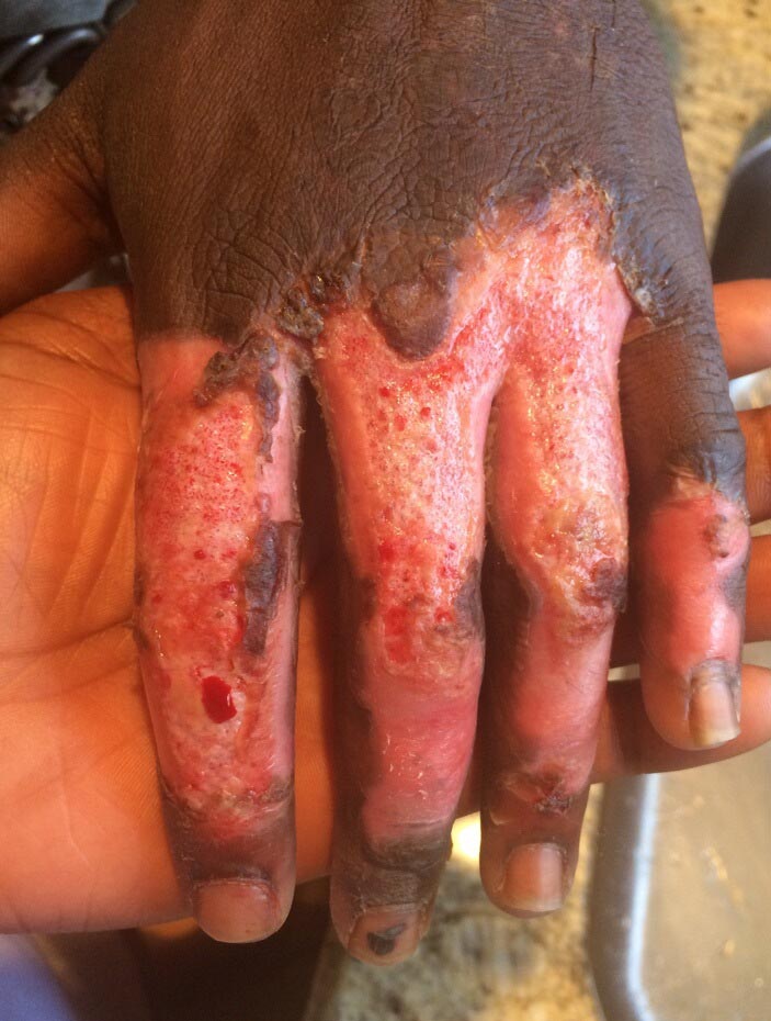
FIGURE 7, WEEK 6
Notice that the pigment is returning to the distial portion of the middle digit where the skin was peeling. Also notict that more skin is peeling and regeneration is occuring in more areas now.
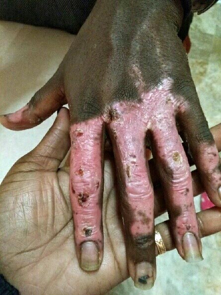
FIGURE 8, WEEK 8
Notice that the white peeling of the skin is a dominant quality at 8 weeks as the skin is being regenerated repigmenting itself.

FIGURE 9
He stopped taking the silver a few months after his hand caught on fire. Therefore, the tissue did not get to finish being regenerated. We are hoping he will begin his routine again to afford Ag the opportunity to finish regenerating the injured tissue.
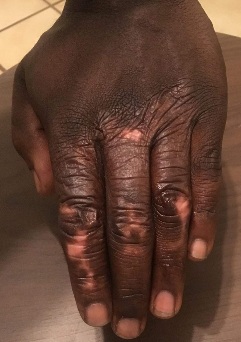
FIGURE 10
The hand below in Figure 9 has already healed to the point of being able to open. The posing of the hand is merely to demonstrate the range of motion that the hand had prior to age 30.

FIGURE 11
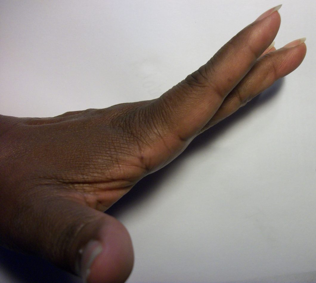
FIGURE 12
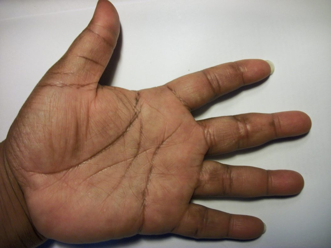
FIGURE 13

FIGURE 14
The finger in figure 14 below, fingerprints are clearly visible though some scar tissue is still visible in the fold of the joint. Hyperpigmentation is still present where some of the old scarring was once present. As colloidal silver is continued to be used, the hyperpigmentation dissipates.
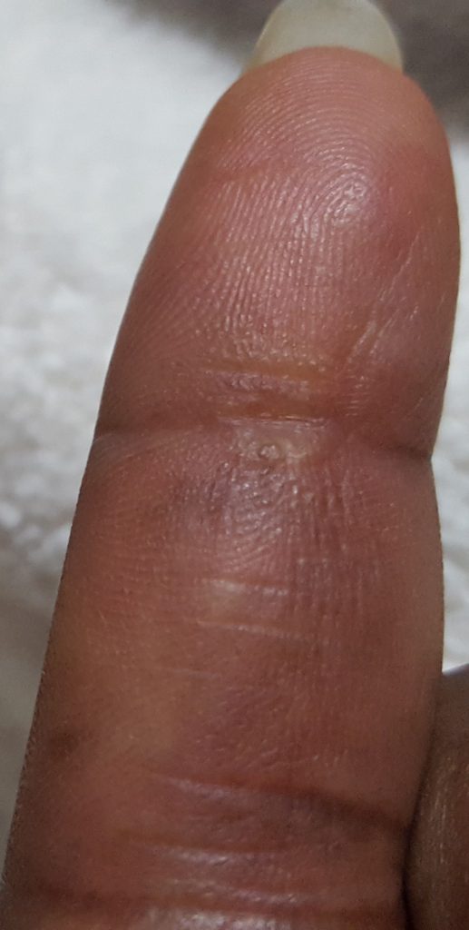
FIGURE 15
Figure 16

FIGURE 17
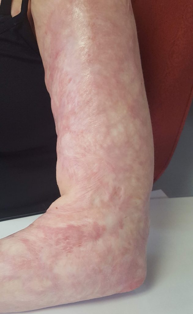
Note that this arm in figure 17 had been healing with vitamin C and colloidal silver for 2 weeks before the picture was taken. Therefore, a lot of the thickness of the scarring had already decreased by the time the picture was taken. There is no hair growing anywhere in the burn areas. Also note the cherry red coloring to the elbow.
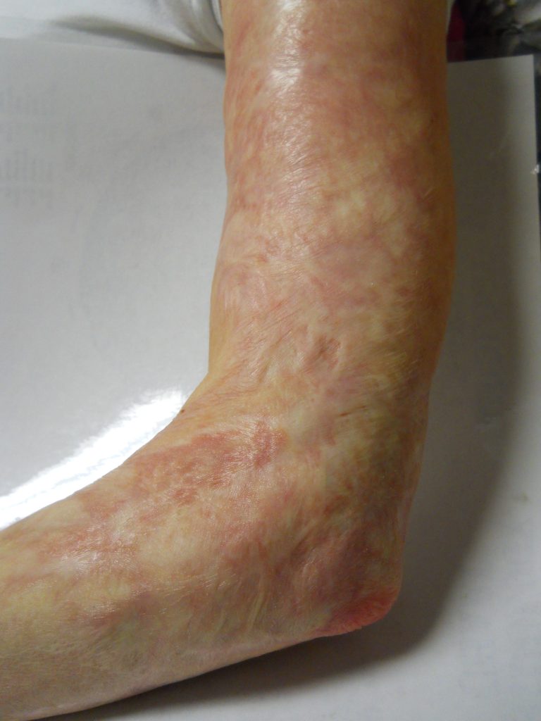
FIGURE 18
Note the thinning, blending and smoothing of the skin. Hair is growing in multiple areas.
FIGURES 19 & 20
The first picture is taken 2 weeks after the CSRBT2019 protocol was initiated while the second is taken after 2 years of being on the CSRBT2019 protocol. Note the thinning, smoothing and blending of the skin. Note the peeling of the skin. Finally, note how the color of the elbow is returning to normal.

FIGURES 21, 22 AND 23
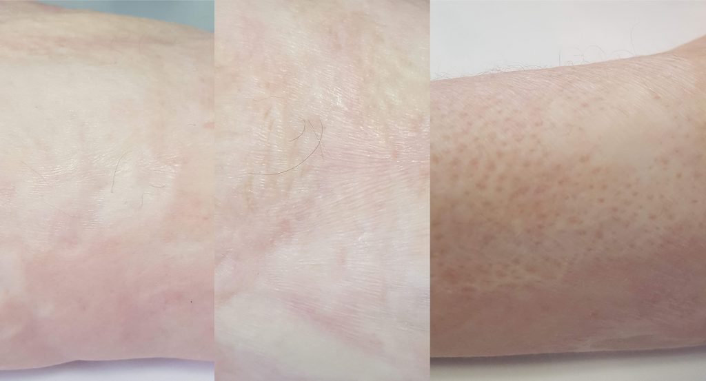
FIGURE 24