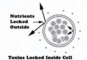
 There are cells in the blood that spend their days gobbling up toxins. Then these cells (called phagocytes) try to break down the toxins into harmless substances and release them. If they can’t break down toxins, such as mercury, they hold onto them. Once filled with toxins, they lock them inside, as shown in the diagram. This prevents the toxins from escaping while the cell tries to deliver them to a new storage site, such as bone marrow, fat, or muscle. In some cases, these cells will hold on to the toxins until they die.
These cells are known to hold onto toxic heavy metals (THMs) in an attempt to keep the blood clean. ([1]) Both Pb and Cd increase phagocytic activity. ([2])
Darkfield Microscopy and Toxins
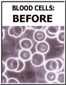 The picture to the right ([3]) is a view of the blood before the dermal chelation occurs. The blood is viewed under darkfield microscopy, a specialized imaging technique that reveals toxins in the blood and blood cells as cloudiness. These toxins can consist of anything that a phagocyte would gobble up.
Dermal Chelation and Toxins
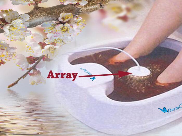 With dermal chelation, the client places their feet into the equipment shown on the left. The equipment is turned on for 1 or more hours. For optimal results, the array (the circular object between the client’s feet) can be changed every 30 minutes. The dermal chelation system delivers a current of 2.1 amps to the water. The current enters the person’s feet and passes throughout their body.
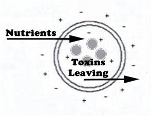 The current will gently unlock the blood cells and other cells, allowing the positively charged toxins locked inside to leave the cells, as seen on the right. Cells with more toxins will release more than cells with barely any toxins stored in them. Then the cells will lock again. The equipment will continue to gently unlock the cells throughout the process, allowing cells storing more toxins to release the most toxins.
Dermal Chelation’s Binding to Toxins
As the current flows through the water in the foot tub to reach the client’s feet, it separates the oxygen and hydrogen from the water molecules, as shown schematically below.

As the H+ gas leaves the water and enters the atmosphere, H2 bubbles can be seen popping on the surface of the water. The HO– ions are magnetized by a magnet in the control panel, acting on one of the wires running to the array between the feet. The magnetized HO– is propelled away from the array, increasing the concentration of magnetized HO– around the feet. The soles of the feet contain many pores that will allow the magnetized HO– to enter. Some of the oxygen entering the pores on the soles of the feet will make its way into the bloodstream, much like transdermal medicines enter the blood.

The magnetized hydroxyl ions (HO-) are negatively charged; they can bind with positively charged substances (X+) in the blood, yielding XO- molecules. When the magnet in the control panel reverses its polarity and pulls the magnetized oxygen back out, anything bound to it gets a free ride out of the body. This is what allows the process to be selective and only remove positively charged substances. The microscopic substances released from the body with the oxygen begin to clump together, with like substances attracting like substances. Eventually, the clumps get big enough to be seen by the naked eye.
The fact that substance X+ can be any metal needing to be removed from the body allows this process to be called dermal chelation. However, one must keep in mind that substance X+ can also be anything that a phagocytic cell will ingest, such as nicotine, cocaine, heroin, methadone, alcohol, by-products of medicines, phosphates, etc.
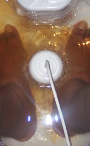
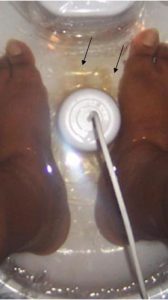
Typically, orange (Or) is the first color to be seen, as in figure 5a and 5b. It is the color seen when the joints are cleansing. The orange from the joints typically appears as ribbons during the first 5 minutes of the dermal chelation process, as seen clearly in both figures. When the joints are being cleansed, you will initially see areas where the water is still completely clear, with no orange color, as the color does not develop uniformly but in ribbons and swirls.
There is an area behind both feet where the water is still clear/uncolored. The orange is darkest near the front of the feet. Note the lack of symmetry. The water in front of the left toes is very orange while the water in front of the right toes is still clear/uncolored. The lack of uniformity between pictures 5a and 5b indicates that the orange represents the cleansed joints. It is thought that the synovial sac is being cleansed. This is consistent with what we have seen in over 8,000 dermal chelations. In over 8,000 dermal chelations, we found an 80% correlation with significant joint issues.
Control Pictures for Dermal Chelation
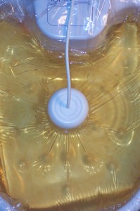
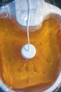
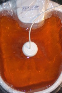
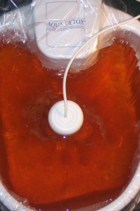
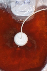
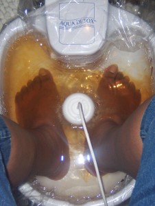
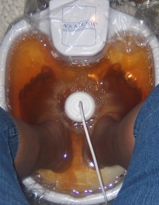
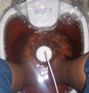
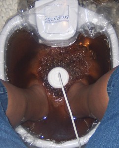
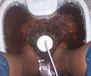
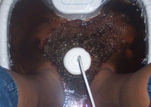
The pictures below are control images showing the dermal chelation system operating with a new array and no feet in the water for 30 minutes. The pictures were taken at 5-minute intervals for the first 30 minutes of the dermal chelation process. Orange is also the color seen when the equipment is operated with a new array and no feet in the water. In this case, the orange color appears uniformly throughout the water, never in ribbons, streams, or swirls as seen in Figure 3. Therefore, a uniform orange color is the control color.
DERMAL CHELATION WATER CONTROL AND RESULTS AT 5 MIN INTERVALS
| 5 MINS | 10 MINS | 15 MINS | 20 MINS | 25 MINS | 30 MINS |
|---|---|---|---|---|---|
 |  |  |  |  |  |
 |  |  |  |  |  |
| 5 MINS | 10 MINS | 15 MINS | 20 MINS | 25 MINS | 30 MINS |
You may need to scroll horizontally to view all of the images in the chart above. As you scroll, note that the result pictures with feet in the water look very different from the control pictures without feet in the water. Though some blackness does appear near the array in the control pictures, it is only the amount you see at approximately 10 minutes with feet in the water. The amount of blackness in the control pictures never reaches the total blackness that you see in the 30-minute result picture with feet in the water.
Dermal Chelation results correlate with each individual’s physicians’ diagnoses and/or history of exposure. Therefore, we conclude that the colors and textures of the resulting dermal chelation water indicate what is happening inside the body.
Dermal Chelation Pictures Explained
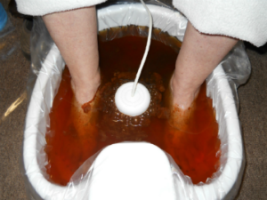 The darkness at the rear of the foot tub in Figure 18 is a black film sitting on the water, thought to be metal removed through the pores of the feet. There is a metallic smell near this film. The orange in the front of the foot tub is caused by the joints (possibly synovial fluid sacs) being cleansed.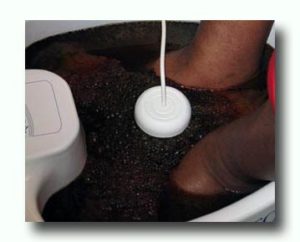
The darkness in the picture on the left, consisting of a film of black metal flecks on the water, was thought to be caused by metal. When the metal turns the top of the water this dark with a thick metallic film, a metallic smell may fill the room. In this particular case, when the water was parted, the darkness was seen all throughout the water as opposed to just floating on top. We suspect that the water’s blackness indicates the liver is being cleansed as well. This correlates with the individual’s diagnosis from her physician. The bubbly foam on top of the water is mixing with mostly black flecks, turning the water black as well.
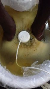 The water resulting from each person’s dermal chelation will look different. As the dermal chelation process progresses, the water will change significantly depending on what the body releases into it. Typically, whatever comes out first gets pushed towards the outer edges of the foot tub as more things continue coming out of the array (the white, circular object in the center of the foot tub). Also, whatever appears on top of the water should be white unless something else is mixing with it as fast as it appears. The more something mixes with it, the more vivid the color will become. Note the flat foam (FF) in the picture to the left, taken five minutes after the process started. It is whiter near the edges of the foot tub. As substances continued to come out of the array, orange and black began to mix with them, changing the color of the flat foam. The black flecks are the metal being removed from the body.
Next:
Dermal Chelation and Dark Cell Microscopy
Resulting Dermal Chelation Water Containing Ammonia, Nitrates, and Phosphates
Understanding Colors and Textures of the Resulting Dermal Chelation Water that Indicates:
Understanding Colors and Textures of the Resulting Dermal Chelation Water that Indicates:
| Bubbly Foam | Dark Green | Blood | Nicotine | Yellow-green |
|---|---|---|---|---|
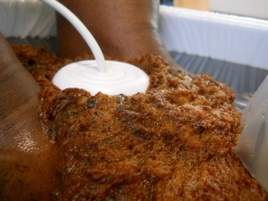 |  | 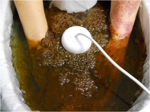 | 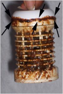 | 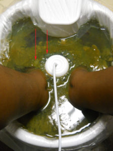 |
You may need to scroll horizontally to view all of the images in the chart above.

Related Topics:
Dermal Chelation For A Total Body Cleanse
Hair Analysis To Determine Your Metal Load
Pictures Of The Water Resulting From Dermal Chelation
Causes Of Heavy Metal Poisoning
Optimum Health’s Concept of Healing
Primary Wellness Consultations
Natural Healthcare Center Location
Email Us: How Can We Assist You?
 Start your path to optimum health by scheduling your appointment today. Member: Certified Natural Health Professionals


References:
[1] Cuha, Elisabete M., et al. “Human & Experimental Toxicology.” Sage Journals, September 1, 2004.
[2] Haschek, Wanda M., Rousseaux, Colin G and Wallig, Matthew A, editors. Haschek and Rousseaux’s Handbook of Toxicologic Pathology. Second edition, Volume 1, Academic Press; 1991,2002.
[3] Aqua Detox. Scientific Research, The Evidence. 2008;8